Description
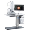
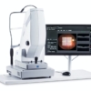
Cirrus Photo 800
One system for Fundus Imaging and OCT
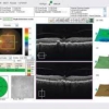
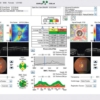
Broader clinical insights, greater diagnostic certainty and added practice value – CIRRUS™ photo from ZEISS delivers all that in a single, integrated system for both fundus imaging and OCT. CIRRUS photo combines a full mydriatic/non-mydriatic fundus camera with proven CIRRUS HD-OCT technology in one compact and highly versatile system. The CIRRUS photo 800 provides multiple insights for comprehensive retina and posterior segment care. Visualizes findings from various modalities. Correlates data from high-density OCT cubes, thickness and layer maps with results from superb color fundus images as well as fundus autofluorescence and fluorescein angiography* images. All in one convenient sitting. Achieve a more comprehensive clinical evaluation. Save time and space. Enhance the examination experience for your patients and staff!
Features:
Broader Clinical Insights
By simultaneously providing high-quality fundus images and OCT scans, CIRRUS photo facilitates broader, more comprehensive diagnostic insights. Each modality by itself is a premier quality diagnostic instrument. Together, they enable you to characterize and examine the patient’s condition more completely and easily.
Greater diagnostic certainty
Comprehensive, high-quality diagnostics form the basis for informed decisions. With its superb multimodality visualizations, CIRRUS photo delivers exceptional insights, supporting greater diagnostic accuracy and certainty.
Added practice value
As a highly efficient and versatile instrument, CIRRUS photo offers substantial value. In addition to streamlining your workflow and
supporting more comprehensive assessments, it saves time and space.
By eliminating the need to move patients to another instrument, it also enhances the examination experience – for patients and practice staff alike.
Specifications:
| Field angle | 45° and 30° |
| Pupil diameter | ≥ 4.0 mm; ≥ 3.3 mm (30° small pupil mode) ≥ 2.0 mm for OCT scans only |
| Refractive error compensation | +35 D … -35 D, continuous |
| Working distance | 40 mm (patient’s eye – front lens) |
| Fixation targets | External and internal |
| Internal | Attention mode and free position or programmed sequences |
| Database | Patient information and images with field angle, FA time, R/L recognition and date of visit are stored |
| Monitor | 23” TFT (1920 x 1200) |
| Instrument table | Asymmetric, suitable for wheelchairs |
| Accessories | Network printer, sliding keyboard shelf, network isolator, FORUM eye care data management system |
Fundus camera
| Capture modes | Color, red-free, blue, red and fundus autofluorescence pictures, as well as pictures of the anterior segment, + fluorescein angiography and ICG angiography |
| Filters | Filters for green, blue and fundus autofluorescence images, UV/IR barrier filters + FA + ICGA: exciter and barrier filters |
| Capture sequence | From 1.5 seconds (depends on flash energy) |
| Capture sensor | CCD 5.0 megapixels |
| Xenon flash lamp | 16 flash levels (30 Ws)-24 flash levels (80 Ws) |
OCT
| Technology | Spectral domain OCT |
| Optical source | Superluminescent diode (SLD), 840 nm |
| Scan speed | 27,000 A-scan per second |
| A-scan depth | 2.0 mm (in tissue), 1024 points |
| Resolution | Axial 5 μm (in tissue), transverse 15 μm (in tissue) |
Computer
| Operating system | Windows Embedded Standard 7 |
| Hard drive | Storage of over 30,000 fundus images with OCT cube scans (present size: 320 GB) |
| Interfaces | USB ports and network connectors, DVI port |
| Export/import | Image formats: BMP, TIFF, JPEG, PNG Patient list, DICOM MWL, DICOM storage |
Dimensions
| Main unit | (W 16.1 x D 18.9 x H 26.8 inches) |
| Weight (main unit) | (72.7 lbs) |
| Rated voltage | 100 … 240 V ±10% |
| Frequency | 50 / 60 Hz |
| Power consumption | 400 VA (w/o instrument table) |
This Zeiss Cirrus Photo 800 comes equipped with:
- 6 Month warranty
- Power table
- Manual
- Dust cover


