Description
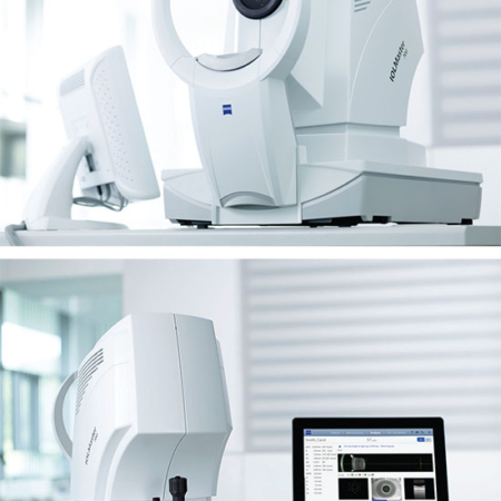
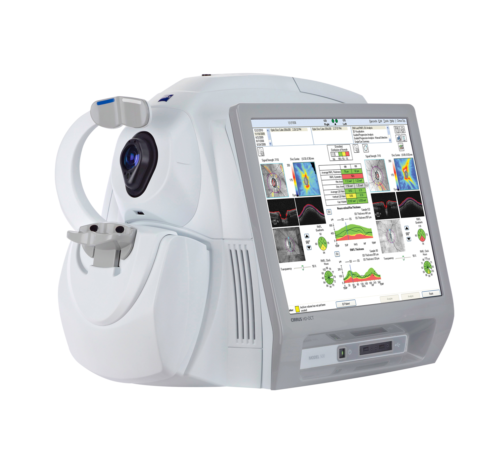
The Zeiss Cirrus HD-OCT 500 provides a great solution for comprehensive ophthalmic practices and offers essential OCT capabilities with a broad range of clinical applications in an easy-to-learn, easy-to-use instrument for the management of glaucoma and retinal disease, retina assessment for cataract surgery, and anterior segment imaging for corneal disease. The new OCT camera enables quick OCT fundus image refresh making patient alignment more efficient. Cirrus HD-OCT is a clinical assessment tool that features industry defining advancements. It is a quick and effective OCT option which allows you to localize services and improve the level of service provision.
Features:
- Macular Thickness Normative Data
- Guided Progression Analysis (GPA™)
- Macular Thickness OU Analysis
- Ganglion Cell Analysis
- Macular Thickness and Change Analysis
Visualization at the speed of CIRRUS
Analyzing a single pathology from multiple views provides comprehensive insight and analysis of the clinical situation. How this helps you:
- Spot small areas of pathology. Tightly spaced B-scans, (either 30 or 47 μm apart), in the cube ensure that small areas of pathology are imaged. For reference, a human hair is about 40-120 μm in diameter.
- Visualize the fovea. Scans that are spaced further apart than in the CIRRUS cube may miss the central fovea.
- Fuel for analysis. Millions of data points from the cube are fed into the Zeiss proprietary algorithms for accurate segmentation, reproducible measurements and registration for change analysis.
- Take the pressure off the operator. As long as the scan is placed in the vicinity of the fovea or optic nerve, the software automatically centers the measurements after the capture.
- See the tissue from different perspectives. View the cube data from all angles, with 3D rendering, OCT fundus images and Advanced Visualization™.
- Future ready. Previously captured CIRRUS cubes can be analyzed using new analyses.
Specifications:
OCT Imaging
- Methodology: Spectral Domain OCT
- Optical source: Superluminesecent diode (SLD), 840 nm
- Scan Speed: 27k-68K A-scans per secound
- A-scan: mm (in tissue), 1204
- Axial resolution: 5 m (in tissue)
- Transverse resolution: 15 m (in tissue)
Fundus Imaging
- Methodology: Line scanning opthalmoscope (LSO)
- Live fundus image: During alignment and during OCT scan
- Optical source: Superluminescent diode (SLD), 750 nm
- Field of view: 36 degrees W x 30 degrees H
- Frame rate: > 20 Hz
- Transverse resolution: 25 m (in tissue)
Iris Imaging
- Methodology: CCD camera
- Resolution: 1280 x 1024
- Live iris image: During alignment
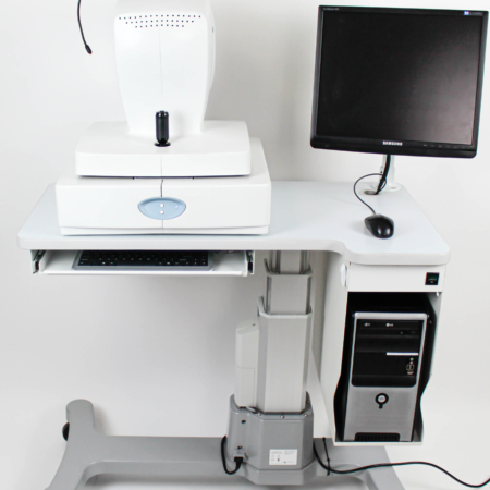
This Zeiss Cirrus HD-OCT 500 comes equipped with:
- 6 Month warranty
- Power table
- Dust cover
- Keyboard and mouse
*Shipping will be padded van service*
*Pictures will be update soon*