Description
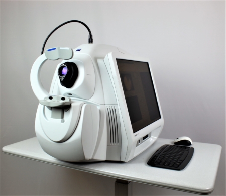
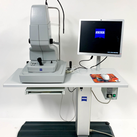
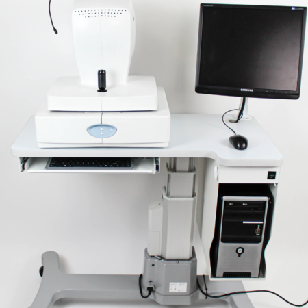
Zeiss Cirrus HD-OCT 5000
Designed for the way you work
The CIRRUS HD-OCT model 5000 is specifically designed to deliver a carefully constructed set of sophisticated applications that build upon one another to deliver rapidly-evolving diagnostics for multiple patient populations. It is a clinical powerhouse with FastTrac, and sophisticated analyses for the busy advanced care practice. Also, The CIRRUS 5000 is the full-spectrum powerhouse OCT/OCTA tailored for busy comprehensive practices. With its growing library of applications, CIRRUS 5000 includes the latest in retina and glaucoma diagnostics, such as OCT angiography and en face imaging, all with the efficiency and reliability to meet the demands of today’s clinical practice.
Features:
- Spectral Domain Imaging
- NEW FastTrac Retinal Tracking System
- NEW Macular Thickness OU Analysis
- Guided Progression Analysis (GPA™)
- Cirrus Cube Analysis
- Macula Normative Data
- Precision Fovea Finder
- HD Smart Scans
- Eye Tracking
- Large 19-inch Monitor
- 3-D Microvascular Imagery
- Acquisition Auto Focus
Features In Detail:
CIRRUS for Retinal Disease:
With the new FastTrac retinal tracking system, precise macular thickness analyses, Fovea Finder, detailed layer maps and more than 100 B-scans at your disposal, CIRRUS provides the framework to comprehensively assess your patient’s retinal condition. CIRRUS data cubes are registered with data from prior visits after the scan is acquired. This enables side-by-side visualization of the same location on the retina for each visit. CIRRUS compares measurements from the current and prior visits to provide a thickness change map that helps you determine next steps for your patient. 3-D cubes and Advanced Visualization can be used for pre-operative planning for VRI disorders. For dry AMD, CIRRUS helps monitor pre-conversion patients today and prepare for tomorrow’s dry AMD therapy.
CIRRUS for Glaucoma:
No other company offers a more complete suite of integrated glaucoma diagnostics. CIRRUS is the HFA’s perfect companion for glaucoma management. Today’s CIRRUS glaucoma applications include RNFL, ONH and angle assessment as well as the new Ganglion Cell Analysis (GCA). When ONH and RNFL status are indeterminate, GCA can sharpen the picture. With CIRRUS angle imaging and central corneal thickness measurements, you have the world’s standard-setting comprehensive tool for glaucoma structural assessment. Guided Progression Analysis (GPA) provides a powerful tool for determining change for RNFL and optic nerve head parameters.
Tracking at The Speed of CIRRUS:
FastTrac reduces eye motion artifacts without sacrificing patient throughput with a proprietary scan acquisition strategy, high speed 20 Hz LSO camera, and single-pass alignment scanning. With FastTrac, scan at the highest resolution at the same location at each visit.
AutoCenter function automatically centers the 3.4 mm diameter peripapillary RNFL calculation circle around the disc for precise placement and repeatable registration. The placement of the circle is not operator dependent. Accuracy, registration and reproducibility are assured.
NEW Smart HD Scan Patterns:
Targeted visualizations of critical anatomy Automatic centering of scans ensures you see the fovea in each patient.
Details Matter — Add flexible HD scans to your macular scanning protocol for an efficient visual assessment of macular status
Get it right the first time — Improves clinic flow by helping to eliminate rescans due to missed fovea
New Smart HD 1 Line scan — Captures and averages 100 b-scan images with automatic centering at the fovea or region of interest. The result is a brilliant image that simultaneously highlights detail in the vitreous, retina, and choroid
CIRRUS 5000 HD-OCT Technical Specifications:
- OCT Imaging: Spectral domain
- Methodology: Superluminescent diode (SLD), 840 nm
- Optical Source: 27,000 – 68,000 A-scans per second
- Scan Speed: 27,000 – 68,000 A-scans per second
- A-scan Depth: 2.0 mm (in tissue), 1024 points
- Axial Resolution: 5 µm (in tissue)
- Transverse Resolution: 15 µm (in tissue)
Fundus Imaging Model 5000
- Methodology Line: Line scanning ophthalmoscope (LSO)
- Live Fundus Image: During alignment and during OCT scan
- Optical source: Superluminescent diode (SLD), 750 nm
- Field of View: 36 degrees (W) x 30 degrees (H)
- Frame Rate: > 20 Hz
- Transverse Resolution: 25 µm (in tissue)
Dimensions/Weight:
- Dimensions of Instrument(L x W x H): 26 x 18 x 21 (in)
- Weight: 80 lbs.
This Zeiss Cirrus 5000 comes equipped with:
- 6 Months Warranty
- Quad-Core Computer with Windows 10 OS
- Latest Software
- Power Table
- Manual and Quick Reference Sheet
- Dust Cover
- Printer *Optional
For more information here is a brochure: https://www.zeiss.com/content/dam/Meditec/downloads/pdf/CIRRUS%20HD-OCT/en-31-010-0014i-cirrushdoct-8.0-ds-ous.pdf

